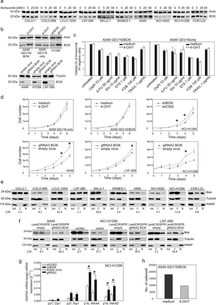
Figure 4.
Stabilization of BOK protein by bortezomib, cell death and proliferation analyses. (a) Cells were treated with bortezomib at the indicated concentration for 24 h; total protein lysates were analyzed by western blotting. (b) Western blot analysis of total protein lysates from A549GEV16/BOK and A549GEV16only cells treated with or without 100 nM 4-OHT for 48h and of H1299, A549 and LXF-289 cells with downregulated BOK using shRNA or CRISPR/Cas9 technology. (c) A549GEV16/BOK or A549GEV16only cells were pretreated with or without 100nM 4-OHT overnight and treated with cisplatin, 5-FU, etoposide, staurosporine, fludarabine or TRAIL for 48 h. Viability was assessed by FITC-AnnexinV/PI staining using flow cytometry. Data represent mean ± S.E.M. from 3–6 independent experiments. (d) Trypan-blue exclusion assay of cell proliferation. Data represent mean ± S.E.M. from 3 independent experiments. Statistical analysis was performed using Student’s t-test. Asterisks indicate a P value lower than 0.05. (e and f) Detection of nuclear BOK by western blot analysis. Nuclear and cytoplasmic fractions were prepared from NSCLC cells. The purity of fractions was confirmed by detection of PARP (nuclear) or Tubulin (cytoplasmic). The cytoplasmic/nuclear ratio of BOK protein expression (C/N) is shown below the blots. (g) mRNA expression of GAPDH-normalized cell cycle regulators by RT-qPCR. Data represent mean ± S.E.M. from 3 independent experiments. Asterisks indicate a P value lower than 0.05 analyzed by Student’s t-test. (h) Anchorage-independent growth of A549GEV16/BOK cells in soft agar for 7 days. Cells were treated as indicated with or without 100 nM 4-OHT.

