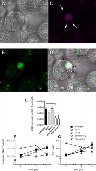
Figure 3.

A–D: Confocal microscopy images of dendritic cells incubated with polymer‐ ImQ conjugates. Murine BMDCs (A) were stained with LysoTracker Green (504 nm/511 nm) for 1 hour (B) and with polymer‐ImQ (390 nm/415 nm). LysoTracker Green accumulates in cellular compartments with low internal pH and stains lysosomes (green, B). Polymer‐ImQ accumulates in lysosomes and in endosomes (C and D, purple regions indicated by arrows). E–F) Conjugates activate T cells and IFN‐γ release. Splenocytes were isolated from transgenic OT‐I mice, labeled with CFSE and stimulated for 4 days with R837, R848 or conjugate, together with the ovalbumin derived SIINFEKL‐peptide, which stimulates ovalbumin specific CD8+T cells from OT‐I mice. On day 4, CD8+T cells were analyzed for CD25 expression and CFSE diluted cells, considered as proliferating T cells by flow cytometry. E) CD25 expression depicted as mean fluorescence intensity (MFI) is shown for splenocytes cultures containing 1 μm conjugate. F) Absolute cell numbers of proliferating CD8+T cells per splenocyte culture were calculated in CFSE diluted dividing T cells using a dosage ranging from 0.1 to 5 μm. G) Supernatants from splenocyte cultures were analyzed for IFN‐γ secretion. Mean ± standard deviation from triplicate analysis is shown for CD25 expression, *) p<0.05, **) p<0.01. Polymer alone and SIINFEKL peptide alone dissolved in DMSO served as negative controls.
