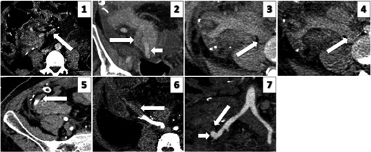
Figure 1.

Representative cross‐sectional images demonstrating grades 1‐3 of venous and arterial pancreas allograft thrombosis. IMAGE 1: Grade 1 venous thrombus: axial image of a portal phase study demonstrating non‐occlusive thrombus (arrow) within an SMV tributary within fat, at the transected margin of the graft. IMAGE 2: Grade 2 venous thrombus: coronal reformatted image of a portal phase study demonstrating non‐occlusive thrombus extending along the splenic vein within the body of graft pancreas (arrow). Adjacent patent splenic artery (short arrow). IMAGE 3: Grade 3 venous thrombus: unenhanced axial image demonstrating acute hyper‐dense thrombus in an expanded SMV. IMAGE 4: Grade 3 venous thrombus: portal phase axial image demonstrating thrombus in the SMV extending into the IVC (arrow). This component is seen as hypo‐dense within otherwise opacified cava. IMAGE 5: Grade 1 arterial thrombus: axial arterial phase image demonstrating minimal thrombus in the distal splenic artery (arrow). IMAGE 6: Grade 2 arterial thrombus: axial arterial phase image demonstrating thrombus extending into the mid SMA. IMAGE 7: Grade 3 arterial thrombus: (image not from current series; obtained from a library) arterial phase coronal reformatted image demonstrating acute occlusive thrombus expanding the Y graft and SMA (arrow). Enhancement of the residual patent proximal Y graft stump (short arrow)
