
Figure 3.
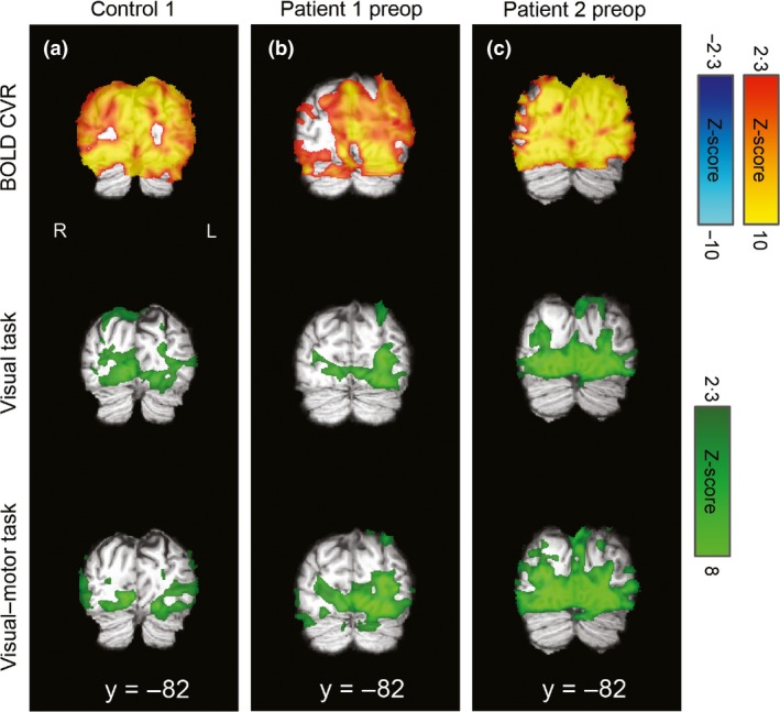
CVR and task‐related activation maps overlaid on the individuals' T1‐weighted images in standard space for visual regions. R=right, L=left, coordinates refer to MNI space. Visual cortex CVR was robustly positive in the illustrative control (a) and patient 1 (b), who both demonstrated bilateral visual activation for both the separate visual task and combined visual–motor task. There were some posterior areas with reduced/absent CVR in patient 2 (c), which corresponded to more lateralized responses, particularly in the visual–motor task, for this patient and ROI (see text and Fig. 1 for details).