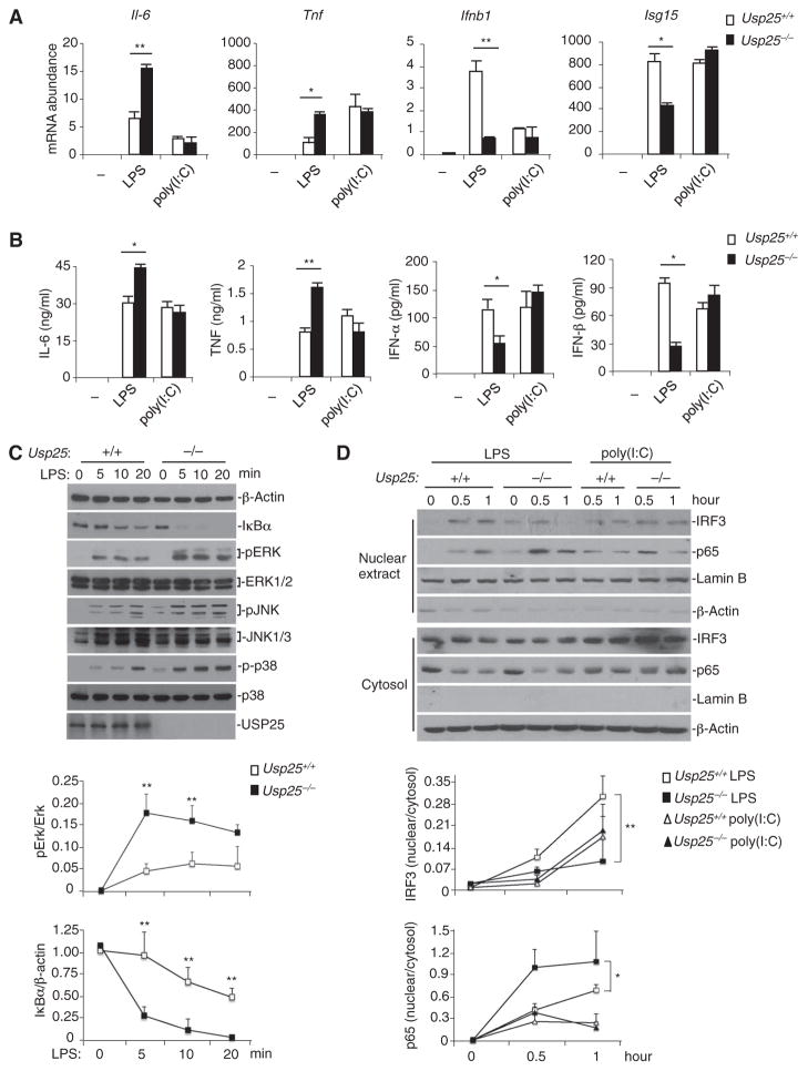
Fig. 4. USP25 restricts TLR4-triggered proinflammatory signaling and promotes TLR4-induced type I IFN signaling.
(A and B) LPS-induced production of proinflammatory cytokines and type I IFNs is differentially regulated by USP25. WT and USP25-deficient BMDCs were stimulated with LPS (0.1 μg/ml) or poly(I:C) (25 μg/ml) (A) for 3 hours before real-time RT-PCR analysis of the abundance of the indicated mRNAs was performed or (B) for 12 hours before ELISA analysis for the indicated proteins was performed. Data are means ± SD from three independent experiments. *P < 0.05; **P < 0.01 by ANOVA. (C) LPS-induced activation of NF-κB and MAPKs is potentiated in USP25-deficient BMDCs. WT and Usp25−/− BMDCs were treated with LPS (10 μg/ml) for the indicated times. The degradation of IκBα and the activation of MAPKs were determined by Western blotting analysis with antibodies against the indicated proteins. (D) LPS-induced nuclear translocation of p65 is enhanced, whereas that of IRF3 is inhibited in USP25-deficient BMDCs. WT and Usp25−/− BMDCs were treated with LPS (10 μg/ml) or poly(I:C) (50 μg/ml) for the indicated times. Nuclear extracts and cytosolic fractions were prepared and analyzed by Western blotting with antibodies against the indicated proteins. Data are representative of at least three independent experiments. The intensities of the indicatedbands in (C) and (D) were quantified, and the ratios of the intensities of the corresponding bands were calculated and are shown in the graphs as means ± SD from three independent experiments. *P < 0.05; **P < 0.01 by ANOVA.

