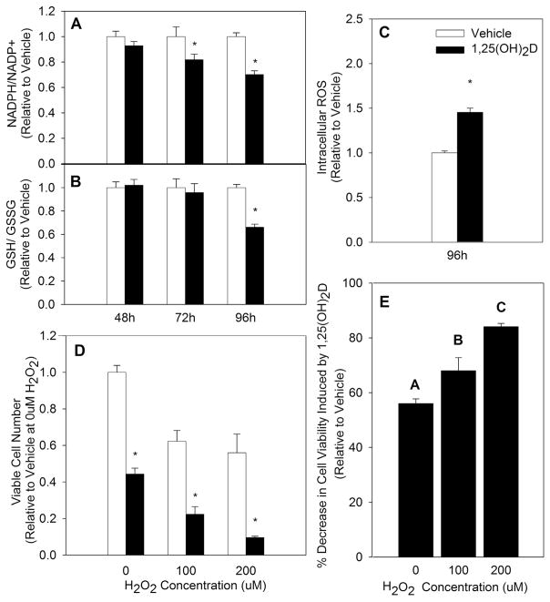
Fig. 1.
Impact of 1,25(OH)2D on Oxidative Stress in MCF10A-ras Epithelial Cells. MCF10A-ras cells were treated with vehicle or 1,25(OH)2D (10 nM). (A&B) The ratios of reduced to oxidized NADPH and GSH were measured after indicated treatment duration and are expressed relative to vehicle treated results of each time point. (C) Intracellular ROS was measured at time indicated and is expressed relative to vehicle treated cells. (D) Cell viability was quantified after a 24 h challenge with indicated H2O2 concentrations following treatment of cells with vehicle or 1,25(OH)2D for 96 h. (E) The percent decrease in cell viability in 1,25(OH)2D-treated MCF10A-ras cells is represented relative to vehicle treated cells at each indicated H2O2 concentration. Values are expressed as mean ± S.E.M. Asterisk indicates significant difference from vehicle (P < 0.05). Bars with different letters are significantly different (P < 0.05).

