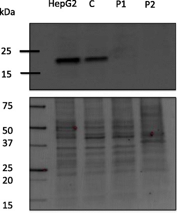
Fig. 1.

Western Blot analysis of TMEM199 protein. Cell layer protein extracts from patient 1 (P1) and patient 2 (P2) fibroblasts were probed for TMEM199 using anti-TMEM199 antibodies (upper gel). An in-house control fibroblast cell line (C) and the commercially available HepG2 hepatocyte cell line were used as controls. The expected position of TMEM199 is at around 19 kDa. Bands detected on the membrane (lower gel) were used as loading controls
