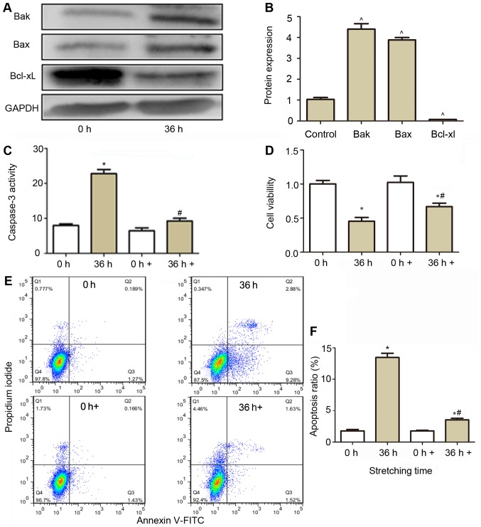
Figure 4.
Effect of caspase-3 inhibitor on the activity of caspase-3, cell viability and apoptosis of CESCs subjected to a 20% stretch at a frequency of 1 Hz for 36 h. (A) Western blot analysis was performed to analyze the expression of Bak, Bax and Bcl-xl. GAPDH was used as a loading control. (B) Quantification of the expression of Bak, Bax and Bcl-xl from (A). (C) Effect of caspase-3 inhibition (0 and 36 h+) on the caspase-3 activity in normal (white column) and stretched cells (brown column). (D) The cell viability was assessed after caspase-3 was inhibited (0 and 36 h+), using a Cell Counting Kit-8 assay in normal (white column) and stretched cells (brown column). (E) Apoptosis was detected using aby flow cytometric Annexin V/PI assay. Representative flow cytometry dot plots of CESCs after double staining with Annexin V-FITC and PI are displayed. (F) Apoptotic indices of CESCs from E. Static CESCs (0 h) were used as a control. +, group incubated with caspase inhibitor Z-VAD-FMK. ^P<0.05 vs. control group; *P<0.05 vs. 0 h group; #P<0.05 vs. 36 h group. CESCs, cartilage endplate-derived stem cells; Bcl-2, B-cell lymphoma 2; FITC, fluorescein isothiocyanate; PI, propidium iodide; Bak, Bcl-2 antagonist killer protein; Bax, Bcl-2-associated X protein; Bcl-xl, Bclextra large protein.

