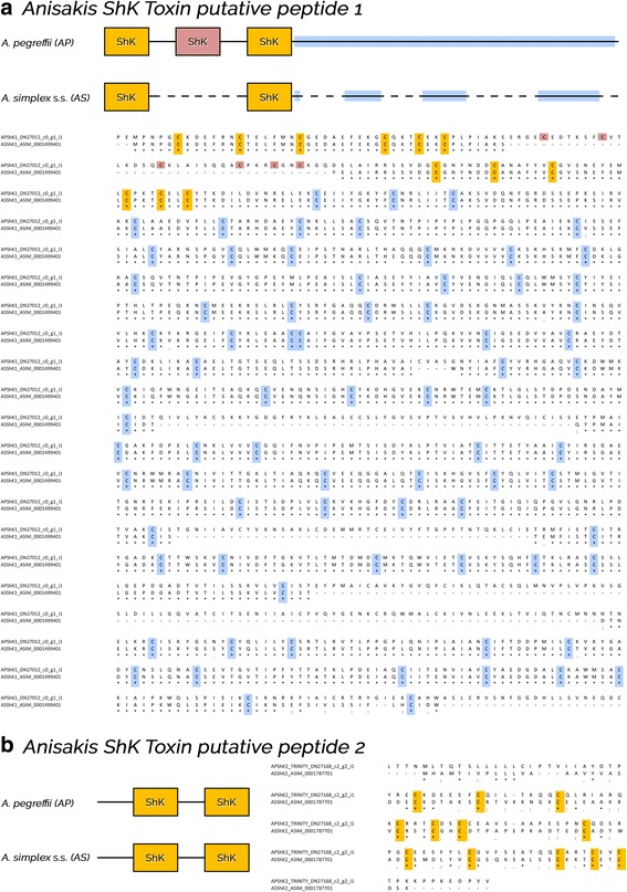
Fig. 3.

Organization and alignment of two ShK toxin putative peptides (a, b) in Anisakis pegreffii (AP) and A. simplex (s.s.) (AS). ShK-cysteines are highlighted in yellow and light red; cysteine-rich domain in light blu. Identical residues, conservative and semi-conservative substitutions are indicated with an asterisk, colon and semicolon, respectively
