Fig. 2.

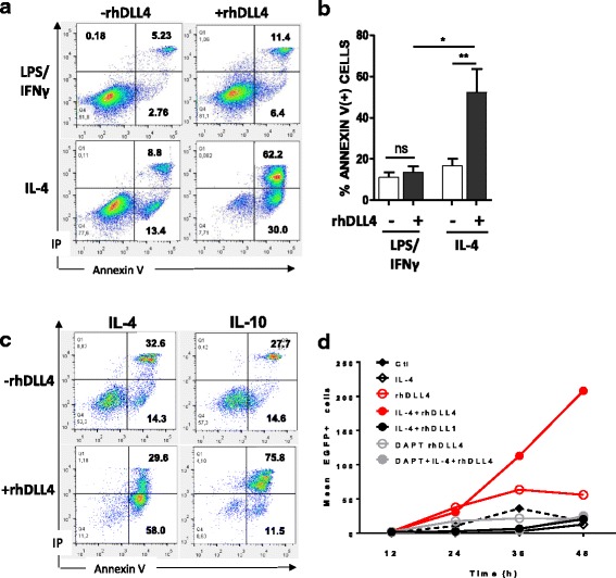
DLL4 triggers specific apoptosis during M2 macrophage polarization. Analysis of apoptosis after M1 (LPS/IFNγ) or M2 (IL-4) polarization with (+rhDLL4) or without (−rhDLL4) coated rhDLL4. a, b, c After 48 h of polarization cells were stained by Annexin V/Propidium Iodide (IP) and the fluorescence was measured by flow cytometry. a Results shown are representative dot plots from one out of 5 independent experiments. b Percentage of Annexin V positive cells among polarized M1 or M2 subsets (means ± SD; n = 5). c Analysis of apoptosis in M2 macrophages polarized with IL-4 or IL-10 with or without coated rhDLL4 (n = 3). Mann-Whitney test was used for statistical analysis of the results (*p < 0.05 and ** if p < 0.01). d Time lapse analysis of caspase 3/7 activity upon M2 polarization. Cells were polarized for 48 h into M2 macrophages (IL-4) or maintain at M0 stage (-IL-4) with or w/o immobilized rhDLL4 or rhDLL1 and with or w/o γ-secretase inhibitor (DAPT). Curves represent the number of apoptotic cells in each condition during the time of the experiment. Data are expressed as means ± SD of EGFP+ cells (activated caspase 3 positive cells) from at least 3 fields per wells
