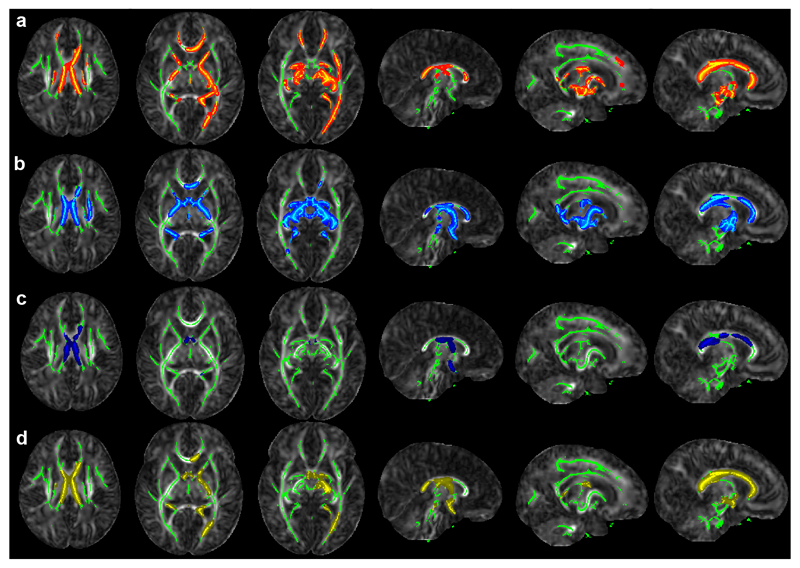
Figure 2.
Clusters in which VP infants differ from LP MCDA twins.
Row a: FA(VP) < FA(LP), Row b: T2(VP) > FA(LP), Row c: MD(VP) > MD(LP), Panel 3: RD(VP) > RD(LP). VP = very pre-term, LP = late pre-term MCDA twins. Colored voxels belong to clusters identified using threshold-free cluster enhancement at the p < 0.05 level using 500 permutations. Green marks the TBSS-demarcated WM skeleton. Results are shown on the JHU-Neonate FA template. Abbreviations used: MCDA = monochorionic diamniotic twin; MD = mean diffusivity; RD = radial diffusivity; WM = white matter; FA = fractional anisotropy.

