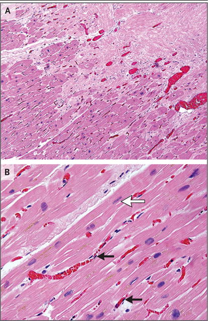
Figure 4. Histologic Assessment of Targeted Myocardium on Autopsy.

Panel A shows prominent small-vessel ectasis at the interface of dense fibrosis (upper right) and viable myocardium (lower left) in postmortem cardiac samples obtained from Patient 5, who had a fatal stroke 3 weeks after treatment. There is no acute myocardial inflammation or acute cellular necrosis. Panel B from the transition region shows occasional rectangular “boxcar” nuclei (white arrow) and hypertrophic cardiomyocytes, which are observed in chronic stages of heart failure. Endothelial cells are normal in appearance (black arrows), showing long, thin, nonreactive nuclei. (Hematoxylin and eosin staining was used in both panels.)
