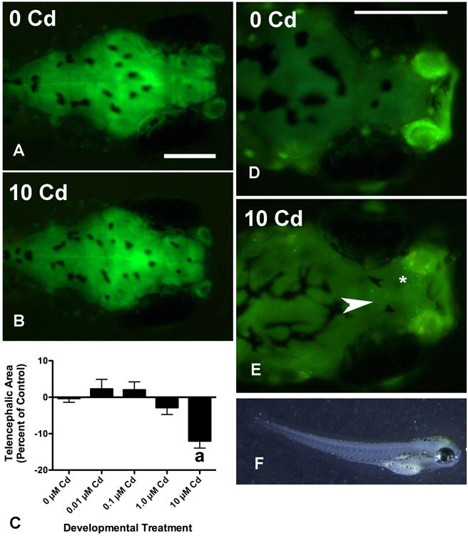
Figure 2. Cd decreases brain size and increases the number of acridine orange-positive (AO+) cells in the forebrain of treated larvae.

Telencephalic area was significantly reduced in larvae treated with 10 μM CdCl2, which is exemplified by comparing the brain in (2A) with that in (2B). While all three gross brain regions were reduced in size at this concentration, the most significant effect was seen for the telencephalon (2C, a: p < 0.001) and hindbrain (not shown, p < 0.001, see Table 1). The data from 6 experiments is shown in Figure 2C and in each of these the telencephalic area of Cd-treated larvae is expressed as a percentage of that from untreated controls in the same experiment (n = 73 for 0 μM, and n = 23-28 for the other concentrations, for statistical analysis the percentages were arcsin transformed). At least some of this effect was probably due to increased cell death as exemplified by comparing AO staining in panels (2D) and (2E). A significant change in the number of AO+ cells was seen at 10 μM of CdCl2 but not at lower concentrations (Table 1, p < 0.001, n = 16 total for each Cd group from 3 experiments). Most of these cells were localized to the olfactory bulbs (asterisk in 2E), however, many were also found in the middle, ventral telencephalon (arrow in 2E). Many 10 μM Cd larvae had an arched appearance (2F). Scale bars in each panel represent 200 μm and the larva shown in 2F is 3.74 mm long.
