
Figure 3. Cd affects heart development and function.
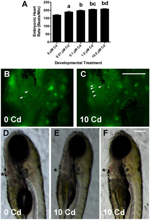
Cd caused a concentration-dependent increase in larval heart rate (3C, data was combined from 3 experiments with a total n = 20 for each Cd group, a: p < 0.01 compared to 0 μM, b: p< 0.001 when compared to 0 μM larvae, c: p < 0.05 when compared to 0.01 μM larvae, and d: p < 0.01 when compared to 0.01 μM larvae). Larvae treated with 10 μM CdCl2 had more AO+ cells (small arrowheads in 2B and 2C) in the heart than untreated larvae. Most 10 μM Cd larvae had hearts that were normal in size and morphology (2D and 2E), but some had an enlarged ventricle and pericardium (3F). The asterisks highlight the ventricles, which showed no obvious defects in looping. There were no discernable differences in morphology or AO+ cells seen in larvae exposed to lower Cd concentrations (not shown). The scale bar for 2B and 2C is 200 μm, and that for 2D-2F is 150 μM.