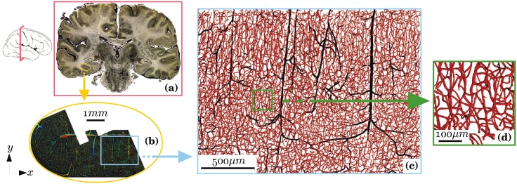
Fig 1. Multiscale architecture of microvascular networks in human brain with a representation of different length scales.
(a) Brain scale (∼63 cm2 × 300 μm): 300 μm-thick cortical section, where blood vessels have been injected with India ink for contrast enhancement [7]. (b) Macroscopic scale (∼18 mm2 × 300 μm): reconstruction of parts of the collateral sulcus by confocal laser microscopy [8]. (c) Mesoscopic scale (∼5 mm2 × 300 μm): region of interest in which vessels of more than 10 μm in diameter are colored in black and vessels of less than 10 μm in diameter are colored in red (diameters have been multiplied by 2 for visualization). In contrast with the capillary bed, the arteriolar and venular trees have a quasi-fractal structure [9]. (d) Microscopic scale (∼0.07 mm2 × 300 μm): detailed view of the capillary bed. The capillary bed is dense and space-filling over a cut-off length of ∼ 50 μm [9].

