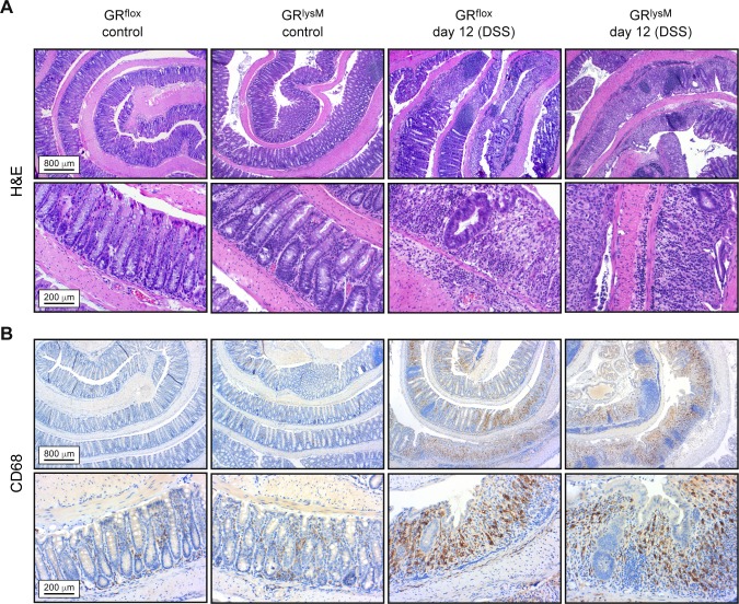
Fig 2. Histological and immunohistochemical stainings of colon sections prepared during the resolution phase of DSS-induced colitis in GRflox and GRlysM mice.
Mice received 2% DSS in the drinking water for 8 days and were sacrificed on day 12. Mice receiving tap water served as controls. The colons were flushed, opened longitudinally and rolled up from distal to proximal to obtain a “swiss-role” for histological analysis. (A) Representative photomicrographs from H&E stained 2 μm colonic tissue sections at 5x (upper panel) or 20x (lower panel) magnification. (B) Representative photomicrographs from 2 μm colonic tissue sections incubated with an anti-CD68 antibody at 5x (upper panel) or 20x (lower panel) magnification. The higher magnification photomicrographs of DSS-treated mice are representative of heavily inflamed areas of the colon. Scale bars correspond to 800 μm and 200 μm, respectively.

