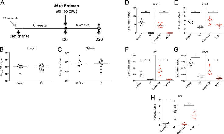
Fig 6. The effect of more severe iron deficiency on susceptibility to M.tb.
Female 4–5 week old C57BL/6 mice were fed an iron deficient (2–6ppm) or control (200ppm) diet for 6 weeks prior to infection with 50–100 CFU of aerosolised M.tb Erdman (A). Mice remained on the respective diet until 4 weeks post-infection, when animals were sacrificed (a total of 10 weeks on their respective diets). Lungs and spleen were harvested for enumeration of CFU (B+C), and livers for gene expression analyses for Hamp1 (D), Fpn1 (E), Id1 (F), Bmp6 (G) and Tfrc (H). Mann-Whitney tests were performed to compare groups where *, **, *** and **** indicate p = <0.05, p = <0.01, p = <0.001 and p = <0.0001, respectively. N = 8 animals in infected groups and n = 6 in uninfected groups. Black symbols represent uninfected animals, red points infected animals except for CFU graphs where all animals are infected. Closed circles represent control animals and open circles, iron deficient animals.

