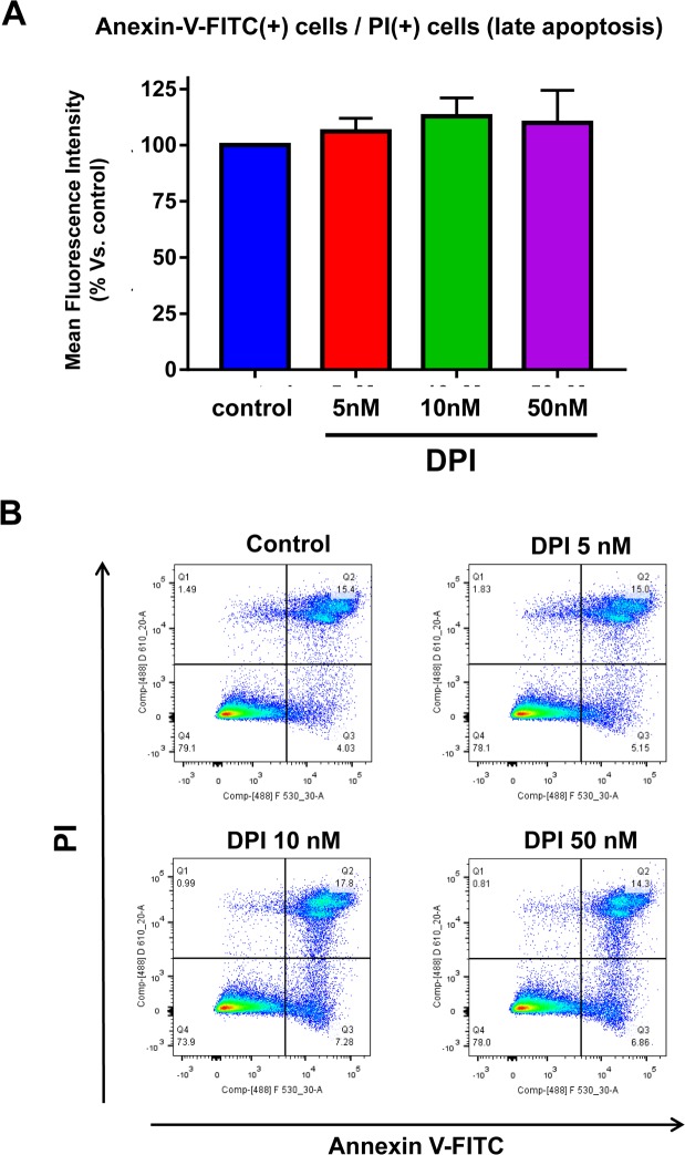
Figure 10. DPI is generally “non-toxic” and does not increase the apoptotic rate in MCF7 cell monolayer cultures.
Briefly, 300,000 MCF7 cells were plated in 6-well plates in complete media supplemented with 10% HiFBS. On the next day, the cells were treated with DPI (5, 10, or 50 nM) for 24 hours. Vehicle alone (DMSO) for control cells were processed in parallel. At least 30,000 events were recorded by FACS using LSRII. The results presented are the average of three biological replicates analyzed in independent experiments and are expressed as mean fluorescence intensity. (A) Bar-graphs are used to summarize the overall results; (B) Representative FACS tracings are also shown. Note that DPI fails to significantly increase the apoptotic rate in MCF7 cell monolayers.

