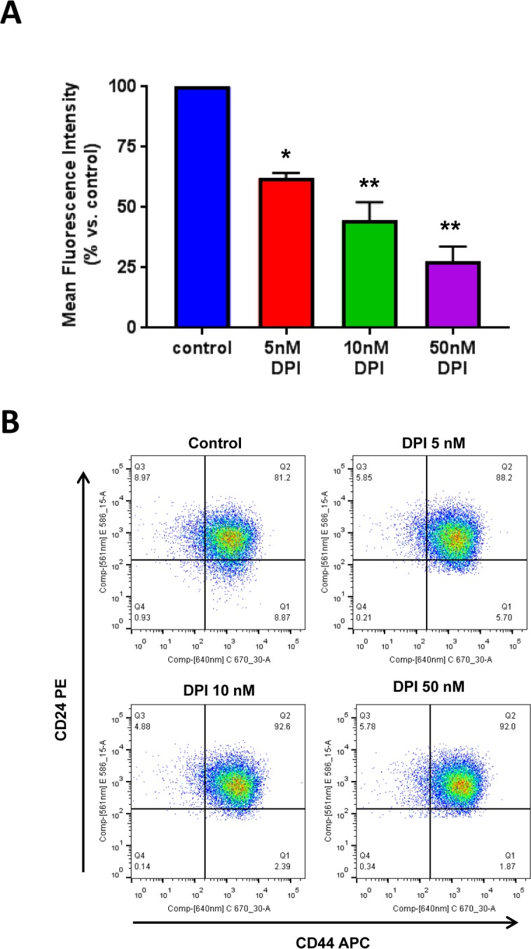
Figure 8. DPI selectively eliminates CSCs from the total cell population.

MCF7 cells were cultured for 5 days as monolayers, in the presence of DPI (5, 10 and 50 nM). Then, the cells were harvested and subjected to FACS analysis to determine the levels of CSC markers. Panel (A) shows that the CD44+/CD24- cell population (which serves as a marker for breast CSCs) is dose-dependently reduced by DPI treatment, with an IC-50 of 10 nM. Panel (B) contains dot plots showing the double fluorescent CD44+/CD24- FACS assay. Note that the signal has significantly decreased in the lower right quadrant (Q1), after DPI treatment. * p<0.05, ** p<0.01.
