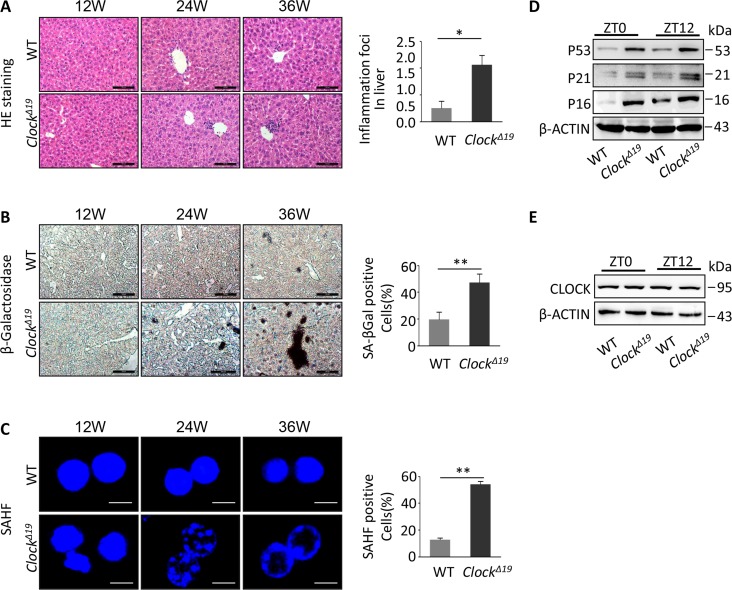
Figure 1. Accelerating the liver aging phenotypes of mice with ClockΔ19.
(A) Histological analysis of WT and ClockΔ19 mice (12, 24 and 36 week old). HE staining of inflammation foci in liver. Data were analyzed by Student's t-test and displayed as the mean ± S.E.M. Asterisks indicate values significantly different from WT (n=4 for all groups). **, P<0.01; *, P<0.05. (B) Liver tissues stained for SA-β-gal activity. The results are expressed as the mean ± S.E.M. A significant increase in the number of positive areas was observed with ClockΔ19 (n=4 for all groups). **, P<0.01; *, P<0.05. (C) Hepatocytes were stained for SAHF foci in WT and ClockΔ19 mice. Note that the livers of ClockΔ19 mice show a clear aging phenotype. The results of the SAHF analysis are representative images of four experiments. **, P<0.01; *, P<0.05. (D) Immunoblots of liver tissue from WT and ClockΔ19 mice at ZT0 and ZT12 for P53, P21 and P16 indicate the accelerated aging phenotypes. β-ACTIN was used as the loading control. **, P < 0.01 and *, P < 0.05 versus control. n=4 mice per group. (E) Immunoblots of ClockΔ19 in mouse liver tissue at ZT0 and ZT12. Note that there was no change in the protein levels in ClockΔ19 mice (n=4). **, P<0.01; *, P<0.05.

