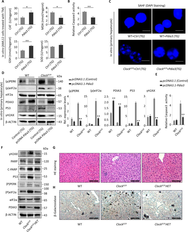
Figure 7. Pdia3 can reverse the aging process of the liver in ClockΔ19 mice.
(A) AML12 cells were transfected with the pcDNA3.1-Pdia3 plasmid (Trans gene, TG) for 24 hours and then stimulated with TM stress (2 μg/mL) for 2 hours. SOD, MDA, GSH and CAT activity were analyzed in the cell homogenate. (n=4) **, P<0.01; *, P<0.05. (B) AML12 cells were treated as described in (A), and the activity of Caspase 3 was measured to reflect apoptotic activity. (n=4) **, P < 0.01 and *, P < 0.05 versus control. (C) SAHF stained via DAPI were visualized by fluorescence microscopy. The primary hepatocytes were extracted, and the number of SAHF was observed after transfection with the pcDNA3.1-Pdia3 plasmid for 24 hours. The SAHF were greatly rescued in ClockΔ19+Pdia3(TG) cells (n=3). (D) The primary liver cells were extracted and transfected with the pcDNA3.1-Pdia3 plasmid for 48 hours. Immunoblotting methods were used to detect the expression of UPR proteins (p)PERK and (p)eIF2α, PDIA3, senescent protein P53, and DNA damage protein γ-H2AX. The statistical results are displayed (right). The results are expressed as the mean ± S.E.M (n=4). **, P < 0.01 and *, P < 0.05. (E) Primary liver cells were extracted and treated as described in (D). Then, the relative activity of Caspase 3 was detected to reflect the level of apoptosis. Note that the apoptotic activity was significantly reduced after transfection with Pdia3. (n=4) **, P<0.01; *, P<0.05. (F) Immunoblots showing the expression levels of γH2AX, PARP, P53, (p)PERK, (p)eIF2a and PDIA3 in the WT, ClockΔ19 and ClockΔ19 heterozygote groups (36 week old). n=3 for all groups. (G-H) Histological analysis of mice as described in (F). HE and SA-β-gal staining of the liver tissue. (n=3 for all groups). Note that the ClockΔ19 HET mice show a decreased inflammation foci and improved aging phenotype.

