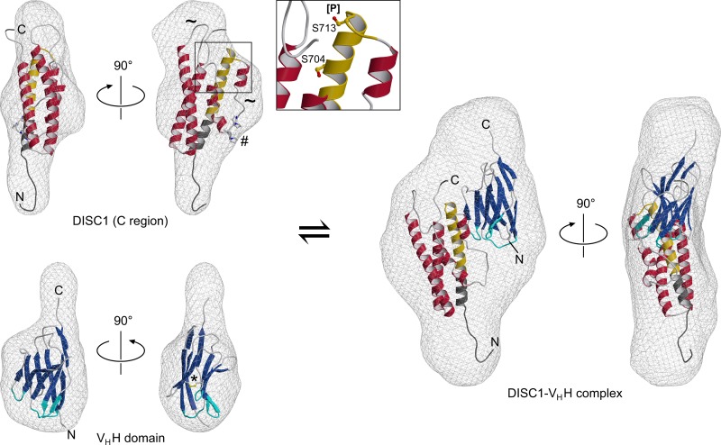
Fig 5. Ab initio SAXS reconstructions and molecular models for the DISC1691-836 protein, the VHH domain, and their complex.
The hypothetical DISC1 model (upper left) features a single domain containing three longer and three shorter α-helices. Similar to the terminal segments, the two extended loops (tilde symbols) are suggested to be poorly ordered. The positions of functionally important residues S704 and S713 are indicated in a close-up view. Also note the proline-rich motif (ball-and-stick representation, marked by a hash), which is predicted to be readily accessible for protein-protein interactions. The 691–715 region, suggested to participate in the epitope for the nanobody, is highlighted in gold while cloning artifacts at the termini are colored dark gray. The model of the VHH antibody (lower left) displays a canonical immunoglobulin fold, including a conserved disulfide bridge (ball-and-stick model, marked by an asterisk). The CDR segments, as defined by the Chothia criteria [60], are highlighted in cyan. Finally, the DISC1691-836 fragment and the nanobody are predicted to interact in a complex (right) featuring a 1:1 stoichiometry.

