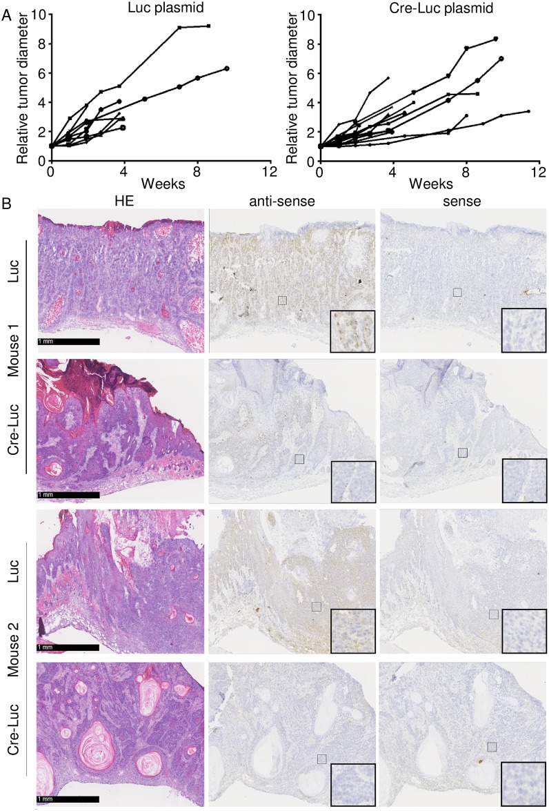
Fig 6. HPV38 E6 and E7 expression is not required for the viability of cancer cells in K14 HPV38 E6/E7 Tg mice.
(A) Electroporated lesions were kept under control and the diameter was recorded weekly. On the day of injection, the lesion diameter varied between 1.2 mm and 2.5 mm for the lesions injected with the Luc plasmid, and between 1.3 mm and 2.6 mm for the lesions injected with the Cre-Luc plasmid. To standardize the measurement, each lesion diameter was set to an arbitrary value of 1 on the day of injection, and the following measurements were adjusted accordingly. The difference in tumour growth between the lesions injected with the Luc plasmid and the lesions injected with the Cre-Luc plasmid was not significant according to an unpaired two-sample Student’s t-test (p = 0.3108, t = 1.052; df = 14). The test was run on data from the fourth week, because afterwards the number of living animals was substantially reduced. (B) Representative images of SCC sections from two different HPV38 E6/E7 Tg mice. Sections were taken from tumours initially electroporated with pS/MARt-Luc plasmid (Luc) or with pS/MARt-Luc-P2A-Cre plasmid (Cre-Luc). The morphological analysis revealed no substantial differences between the specimens; the tumours were all classified as invasive cSCC, with deep penetration into the dermis or into the muscular fibres, and clear and diffuse atypia. The loss of the viral mRNA in the tumours injected with the Cre-Luc plasmid was confirmed by in situ RNA hybridization using a complementary (antisense) riboprobe, while the staining with a sense probe confirmed the specificity of the signal.

