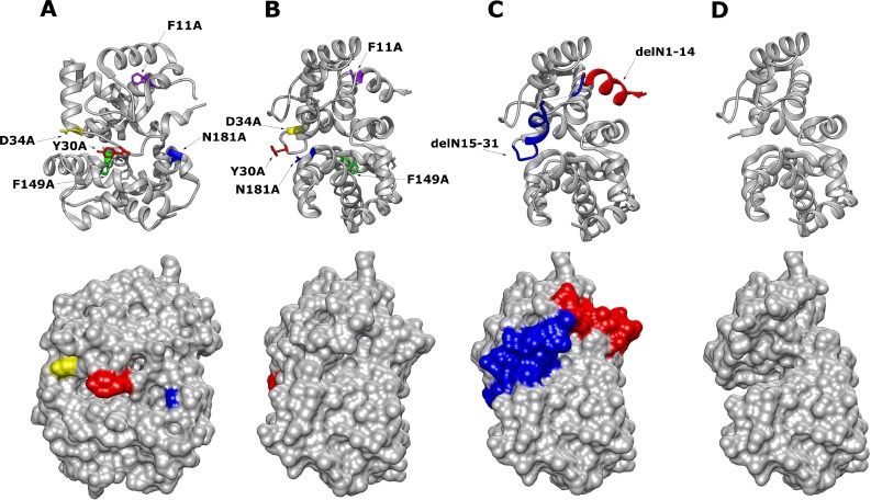
Fig 2. Chimera 3D Model of RVFV N protein monomeric structure with mutated residues highlighted (PDB: 3LYF) [18].
(A) RVFV N protein with highlighted point mutations (top image) and corresponding surface view (lower image). (B) Alternative orientation of the RVFV N protein point mutations at a 90-degree offset to (A). (C) N protein with highlighted N-terminal arm position 1–14 in red, 15–31 in blue. (D) Mutant RVFV N protein with the full delN1-31 arm removed and surface view.

