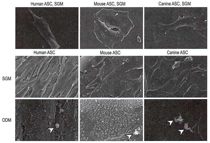
Fig. 1.
Scanning electron microscopy comparing human, mouse, and canine adipose-derived stromal cells. (Above and center rows) Cells were allowed to grow for 7 days in standard growth medium and imaged by scanning electron microscopy technology at 1000× (above) and 600× (center row) magnification. Mouse cells were observed to have a spread and rounded appearance. Human cells were tightly aligned in a thin and elongated pattern. Canine cells were somewhere in between that of mouse and human cells in morphology, with a more fibroblastic appearance than mouse but less spindle-shaped than human cells. (Below) Cells were allowed to grow in osteogenic differentiation medium (ODM) and were imaged by scanning electron microscopy technology at 600× magnification. Under osteogenic conditions, human, mouse, and canine adipose-derived stromal cells adopted intricate cell surface features, including a studding of the cell surface with numerous, small, rounded projections representing bone nodules (arrowheads).

