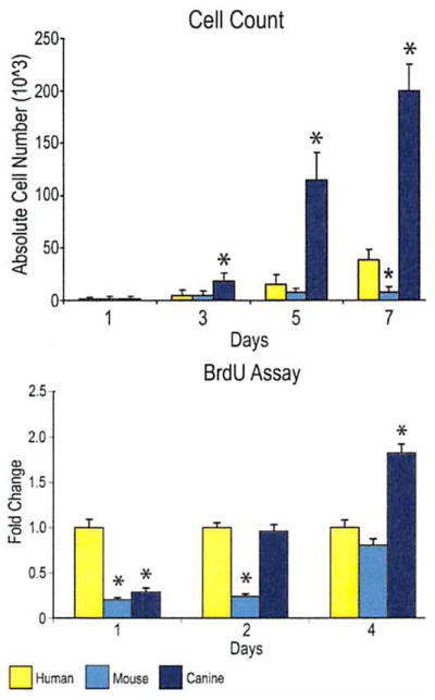
Fig. 2.
Cellular proliferation among human, mouse, and canine adipose-derived stromal cells. (Above) Cell counting assays were performed over 7 days in standard growth medium (Dulbecco’s Modified Eagle Medium, 10% fetal bovine serum) by trypsinization and hemocytometry. (Below) Bromodeoxyuridine (BrdU) incorporation assays were performed over 4 days in standard growth medium by enzyme-linked immunosorbent assay. Labeling reagent was applied for 8 hours in culture (n = 3 for cell counting, n = 6 for bromodeoxyuridine) (*p < 0.05).

