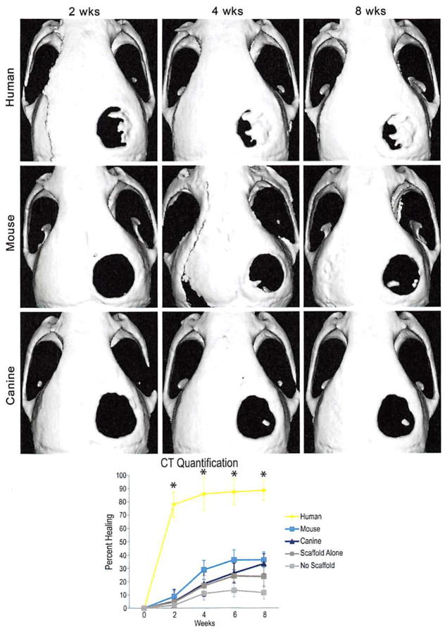
Fig. 5.
(Above) Micro–computed tomographic scans of live mouse calvariae after a 4-mm parietal defect and placement of a 4-mm hydroxyapatite coated poly(lactic-co-glycolic acid) scaffold with 150,000 human (first row), mouse (second row), or canine adipose-derived stromal ceils (last row). Human cells demonstrate a significant amount of healing as early as 2 weeks and nearly complete healing by 8 weeks; mouse cells demonstrate some osseous healing by 4 weeks but never completely heal. Canine cells demonstrate minimal healing around the edges of the defect. (Below) Quantification of mean calvarial healing calculated by dividing the size of the defect at each time point by the size of the defect at day 0 of injury. Human adipose-derived stromal cells have significantly greater healing at all time points (n = 4 mice per group; *p < 0.05). CT, computed tomography.

