Fig. 6.
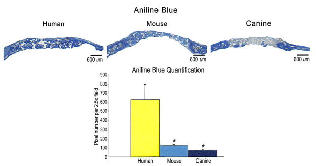
(Above) Histology of calvarial defects. Four-millimeter calvarial defects were allowed to heal for 8 weeks before histologic analysis by aniline blue stain. Pictures were taken of the midpoint of the defect site. In aniline blue stains, bone appears dark blue. (Below) At 8 weeks, aniline blue–positive bone per 2.5 × field was quantified (n = 50 slides per group, n = 5 animals per group). A one-factor analysis of variance was used, followed by a post hoc t test to assess significance (*p < 0.05).

