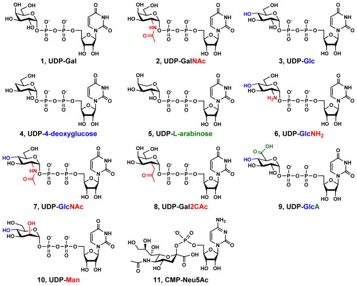
Figure 3.
Structures of UDP-Gal (1), UDP-GalNAc (2), UDP-Glc (3), UDP-4-deoxyglucose (4), UDP-L-arabinose (5), UDP-GlcNH2 (6), UDP-GlcNAc (7), UDP-Gal2CAc (8), UDP-GlcA (9), UDP-Man (10), and CMP-Neu5Ac (11). The structural features that differ other UDP-sugars from UDP-Gal are highlighted in red, blue, and green at C2, C4, and C6 of the Gal in UDP-Gal, respectively.

