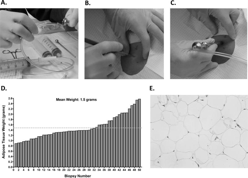
Figure 1. Biopsy Method With the Bergström Biopsy Needle.

A. Tubing was cut at a 45-degree angle in order to fit more securely into the top of the Bergström biopsy needle for generating suction. Tubing inserted into the cutting trochar of the Bergström biopsy needle. B. A 6–7 mm incision was made through the skin up to the hub of a number 11 Bard Parker blade. C. When US guidance was not used, the Bergström biopsy needle was inserted approximately 1.5 inches in the incision site. D. The average SAT biopsy weight taken was 1.5±0.4 grams. E. Representative hematoxylin and eosin staining showing integrity of adipose tissue is maintained when using the Bergström biopsy needle.
