
Figure 1.
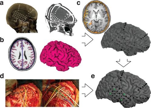
Pipeline Overview. (a) Electrodes localized on postoperative CT. (b) Pial surface and smoothed pial surface created from preoperative MRI. (c) CT‐MRI registration results in electrodes localized beneath the surface; the surface has been made translucent. (d) Intra‐operative photographs show cortical anatomy and electrode placement, allowing accurate identification of anchor points. (e) Result of our localization algorithm as guided by anchor points [Color figure can be viewed at http://wileyonlinelibrary.com]