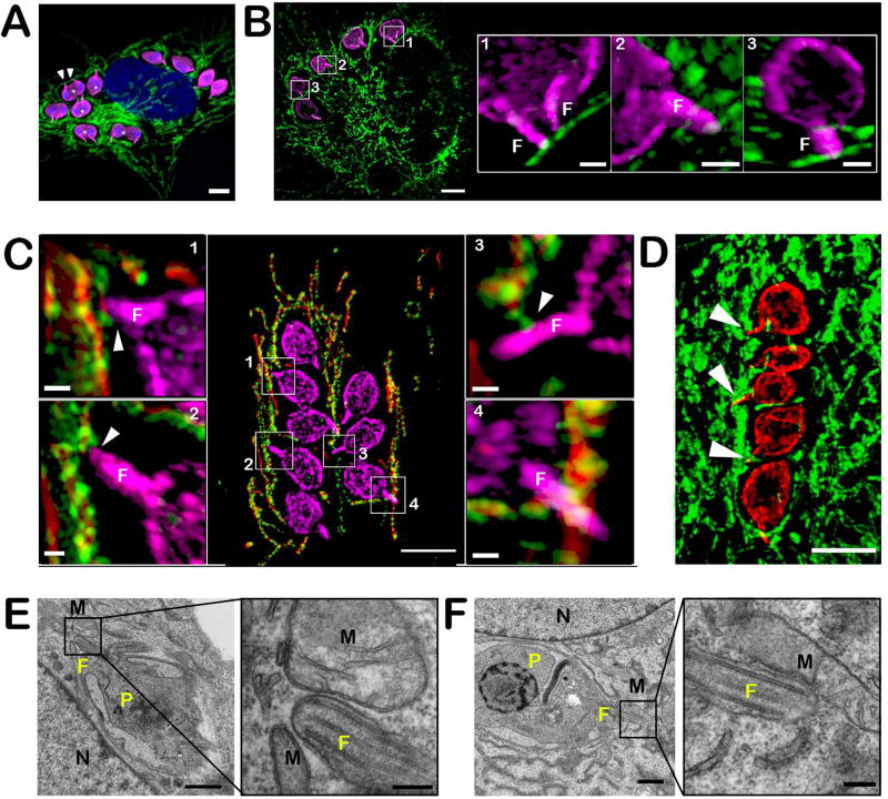
Figure 3. Close interaction between T. cruzi amastigotes and host mitochondria is mediated by the parasite flagellum.
A. Immunofluorescence detection of T. cruzi amastigotes using anti-FCaBP (pink) and host mitochondrial network with anti-ATP5B antibody (green) in infected NHDF (48 hpi). DNA is stained with DAPI. Scale bar = 5 µm. Example of parasite flagella (white arrowheads). B. Super-resolution (SR-SIM) 3D projection of intracellular T. cruzi amastigotes in NHDF 6 hour post-replating following trypsin treatment (42 hpi). Parasites are stained with anti-FCaBP (pink) and the mitochondria is stained with ATP5B antibodies (green). Scale bar = 5 µm. Insets; 3D SR-SIM reconstruction of the framed areas using the 3D projection mode showing the zone of contact between the parasite flagellum (F) and the host mitochondria. Scale bar = 1 µm. C. Super-resolution (SR-SIM) 3D projection showing infected mito-mCherry NHDF. Parasites are stained with anti-FCaBP (pink) and the host mitochondria expressing mCherry fluorescent protein (red) are stained with TOM20 antibodies (green). Scale bar = 5 µm. Insets; 3D SR-SIM reconstruction of the framed area using the 3D projection mode showing the zone of contact between the parasite flagellum (F) and the host mitochondria indicated by white arrowheads. Scale bar = 0.5 µm. D. Super-resolution (SR-SIM) 3D projection of an infected human cardiomyocyte (iPSC-derived). Mitochondria are visualized using an anti-ATP5B antibody (green) and parasites are stained with an anti-FCaBP antibody (red). Flagella in contact with the host mitochondria are indicated by white arrowheads. Scale bar = 5 µm. E and F. TEM of T. cruzi intracellular amastigotes in NHDF cells (flat embedding). P, parasite; N, host nucleus; F, flagellum; M, mitochondria. Scale bar = 1 µm. Insets show the proximity between the amastigotes flagellum and the host mitochondria at high magnification. Scale bar = 200nm.

