Figure 1.
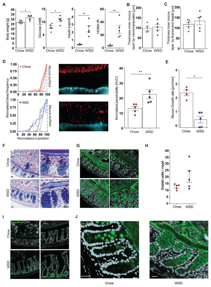
Western style diet feeding impairs the inner colonic mucus layer in mice. Mice (4 – 5 mice/group) were fed a Western style diet (WSD) or chow diet for 8 weeks and metabolic parameters such as body weight, fasting blood glucose and insulin concentration were measured and HOMA-IR was calculated (A). Thickness of the inner colonic mucus layer was measured ex vivo with a micromanipulator by measuring the distance between black 10 μm beads and the epithelial surface (B) or with a confocal microscope by measuring the distance of fluorescent 1 μm (bacteria sized) beads and the stained epithelium (C). (D) Confocal z-stacks, (left), calculated from the position of fluorescent 1 μm beads (center), were used to determine penetrability of the inner colonic mucus layer (right). The median z-stack is shown for each mouse. Fluorescent beads, inner mucus layer and colonic epithelium are indicated in the figure. Turquoise: colonic tissue; red: bacteria-sized beads. (E) Growth rate of the inner colonic mucus layer. (F) Carnoy fixed colonic tissue sections were stained with Alcian Blue/periodic acid-Schiff or with polyclonal antibodies against glycosylated (mature) Muc2 (Muc2-C3, green)(G). Please note that the outer mucus layer is less defined and often not observed in fixed tissue sections. (H) Goblet cell number per colonic crypts. (I) Carnoy fixed colonic tissue sections were incubated with polyclonal antibodies against non-O-glycosylated (immature) Muc2 (Muc2-PH497, green) and glycosylated (mature) Muc2 (Muc2-C3, green) (J). Arrows in (J) indicate mucus secretion inside the crypts. Data in A, D, E H are presented as mean ± SEM. Statistical significance was determined by Mann-Whitney U test with (*) = p<0.05 and (**) = p<0.01. Size bar = 25 μm, representative images are shown. See also Figure S1.

