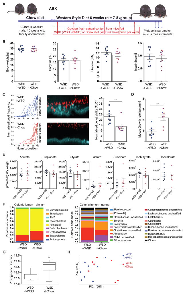
Figure 5.
Microbiota transplant from chow-fed mice prevents deterioration of the colonic mucus layer upon WSD feeding. (A) 10 week old mice (n= 7–8 mice/group) were treated with an antibiotic cocktail (ABX: ampicillin 1 g/l, metronidazole 1 g/l, vancomycin 0.5 g/l, neomycin 0.5 g/l) for 3 days and subsequently switched to a WSD. Mice received a weekly microbiota transplant either from WSD-fed donor mice (WSD->WSD) or from chow-fed donor mice (WSD->Chow). After six weeks metabolic parameter were determined (B). (C) Confocal z-stacks (left) calculated from the position of fluorescent 1 μm beads (center) were used to determine penetrability of the inner colonic mucus layer (right). The median z-stack is shown for each mouse. Turquoise: colonic tissue; red: bacteria-sized beads. (D) Growth rate of the inner colonic mucus layer. (E) Caecal fermentation products of WSD->WSD and WSD->Chow mice. Data in B-E are presented as mean ± SEM. Statistical significance was determined by unpaired t-test for normally distributed data and Mann-Whitney U test for not-normally distributed data with (*) = p<0.05 and (**) = p<0.01. (F) Relative abundance of microbial taxa was determined by 16S rDNA analysis of colonic bacteria on phylum and genus level, for which abundances >1% are shown. (G) Alpha diversity and beta-diversity (weighted Unifrac, H) for the colonic microbial community. Statistical significance for diversity analyses was calculated as described in the methods sections. See also Figure S3 and Table S1.

