Figure 1.
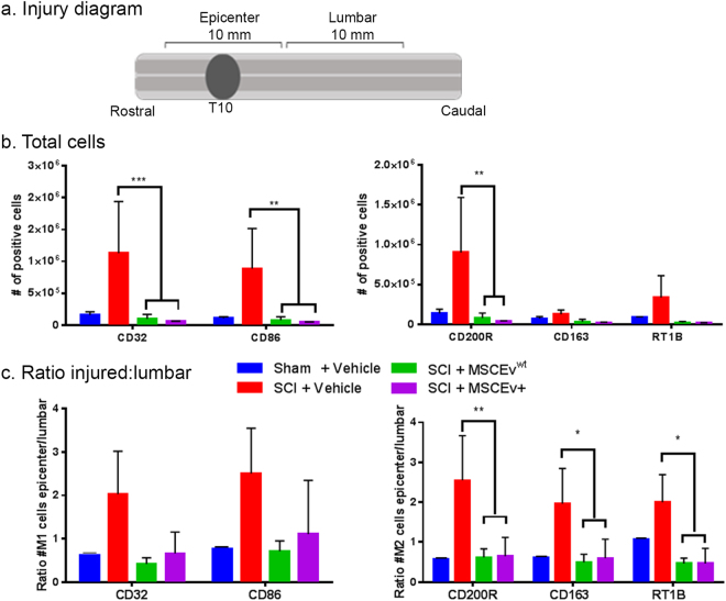
Microglia activation in injured spinal cord Samples of spinal cord tissue containing the injury epicenter (a) were analyzed by flow cytometry for microglia activation state phenotypes. Interestingly, the total number of cells positive for markers associated with pro-inflammatory, M1 microglia (CD32 and CD86) were significantly decreased after treatment with MSCEvwt and MSCEv+ (***p < 0.001, **p < 0.01, respectively)(b). Additionally, lumbar sections of spinal cord were analyzed, revealing the ratio of positive cells in injured sections of spinal cord and distal lumbar sections of spinal cord. Cells positive for M1 associated markers CD32 and CD86 are increased after SCI but treatment with either MSCEvwt or MSCEv+ decrease levels of these cells to near sham values. Cells positive for M2 associated markers CD200R, CD163 and RT1B are significantly decreased (**p < 0.01, *p < 0.05, *p < 0.05, respectively) with treatment of either MSCEvwt or MSCEv+ after SCI. Overall, there is a significant reduction in the number of activated microglia after treatment with either MSCEvwt or MSCEv+, indicating that both EVs are effective in reducing inflammation in the injury epicenter as early as 14 days post-injury (c). Sham + Vehicle, n = 2, SCI + Vehicle, n = 2, SCI + MSCEvwt, n = 3, SCI + MSCEv+, n = 3.

