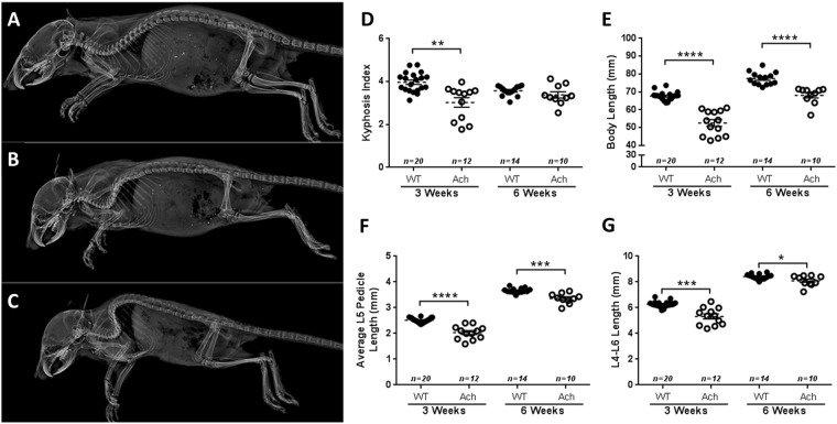
Figure 3.
Axial skeletal defects in Ach mice. Lateral X-rays of 3-week-old WT (A) and Ach mice with mild (B) to severe (C) kyphosis. Kyphosis index (D) and body lengths (E) are shown for WT and Ach mice at 3 weeks and 6 weeks of age. Average L5 pedicle lengths (F) and L4-L6 lengths (G) are shown for the 3-week and 6-week-old mice. The graphical data are represented as mean ± SEM (*0.01 < p < 0.05, ***0.0001 < p < 0.001, ****p < 0.0001).

