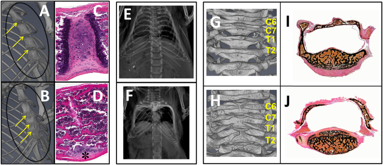
Figure 4.
Abnormalities of sternebrae and vertebrae in Ach mice. 3D micro-CT of sternum with yellow arrows indicating junctions of adjacent sternebrae in representative 3-week-old WT (A) and Ach (B) mice. Corresponding H&E stained sections showing 3-week-old WT mouse (C) with joint cartilage and growth plate and Ach mouse (D) with mature bone replacing the intersternebral joint and growth plates. The asterisk shows receding joint cartilage. X-ray images are shown for the rib cages from representative WT (E) and Ach (F) mice at 3-weeks of age. Note lateral displacement of ribs giving rise to box-shape rib cage in Ach mouse. 3D micro-CT of vertebrae in representative 3-week-old WT (G) and Ach (H) mice reveal incompletely closed dorsal arch and lack of spinous process in Ach mouse. Corresponding von Kossa method stained sections show trabecular bone on the dorsal aspect of thoracic vertebra in WT mouse (I) and fibrous connective tissue instead of bone forming the dorsal arch in Ach mouse (J).

