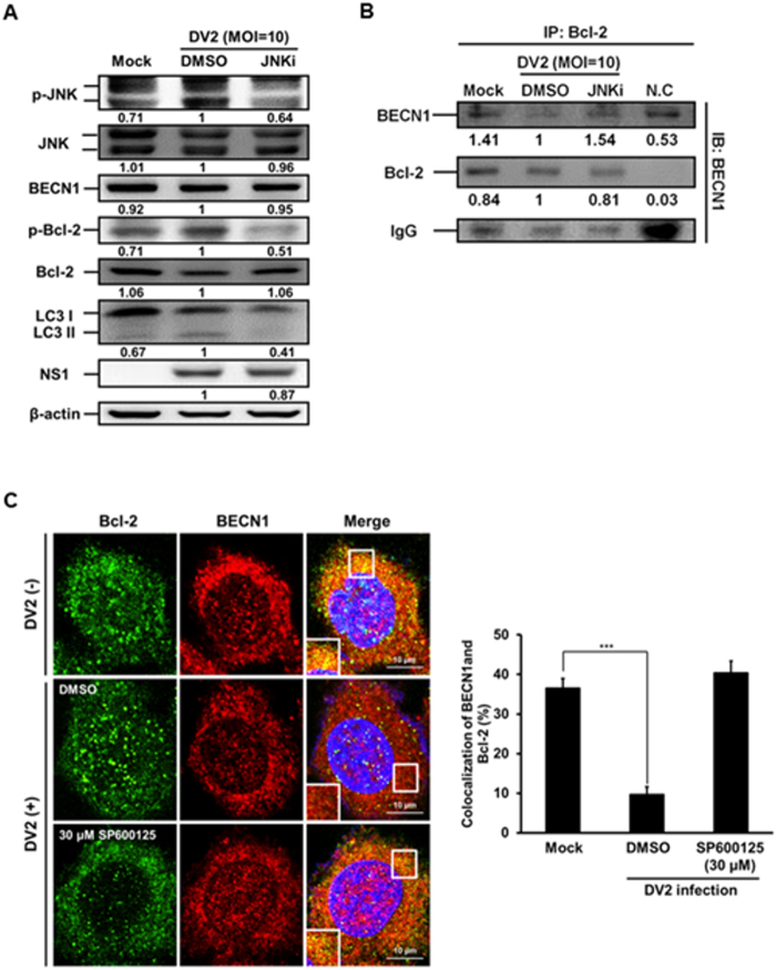
Figure 6.
The phosphorylated JNK leads to decreased interaction between Bcl-2 and BECN1 during DENV2 infection, and the effect was rescued by SP600125 treatment. (A) Huh7 cells were infected with DENV2 (MOI = 10) with or without the treatment of JNKi-SP600125 (30 μM), and the cells were collected at 36 h p.i. The protein levels of p-JNK, JNK, BECN1, p-Bcl-2, Bcl-2, LC3-II/ LC3-I and NS1 were determined by Western blotting. (B) The mouse anti-Bcl-2 antibody (2 μg/ml) was used as the primary antibody to pull-down Bcl-2 protein and incubated with BECN1 antibody at 4 °C for overnight followed by Western blotting to detect the expression of BECN1, Bcl-2 and IgG. The number under the band is the quantification of band intensity and normalized with β-actin. (C) Huh7 cells were infected with DENV2 and treated with SP600125 (30 μM), then the cells were fixed at 36 h p.i. The FITC-labeled Bcl-2 and PE-labeled BECN1 were detected under the confocal microscopy. The relative percentage of colocalization of Bcl-2 and BECN1 during DENV2 infection with or without SP600125 treatment was determined.

