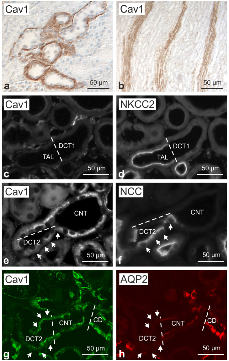
Figure 1.
Renal distribution of caveolin 1 in wild-type mouse kidney. (a,b) Representative bright-field images showing basolateral caveolin 1 (Cav1) distribution along renal distal epithelia (a) as well as a Cav1 signal in medullary vasculature (b); immunoperoxidase/hematoxylin staining, interference contrast. (c,d) Double immunofluorescence staining for Cav1 and Na-K-2Cl cotransporter (NKCC2) shows the transition between NKCC2-positive thick ascending limb (TAL) and NKCC2-negative early distal convoluted tubule (DCT1; bar); TAL and DCT1 are Cav1-negative. (e,f) Immunofluorescence staining for Cav and, on a consecutive section, for DCT-specific Na-Cl cotransporter (NCC) showing the transition between NCC-positive late DCT (DCT2) and ensuing NCC-negative connecting tubule (CNT); moderate basolateral Cav1 signal is detected in DCT2 (arrows); stronger Cav1 signal is observed in CNT principal cells. (g,h) Double immunofluorescence staining for Cav1 and aquaporin 2 (AQP2) shows a moderate Cav1 signal in AQP2-negative DCT2 (arrows) and a stronger Cav1 signal in CNT and collecting duct (CD) principal cells (bars indicate DCT2/CNT and CNT/CD transitions).

