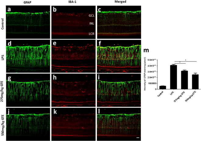
Figure 3.
Green tea extract attenuated the activation of Müller glial cells. (a–c) GFAP staining labelled a dense layer of astrocytes and a few Müller glia processes that run through the whole thickness of the retina in control group. (b) IBA-1 staining showed the localization of a few microglia in the ganglion cell layer (GCL) and inner nuclear layer (INL). (c–f) After LPS insult, the expression of GFAP in Müller glia cells was elevated obviously, which accompanied by an increase in microglial cells in GCL and INL. (g–l) GTE treatments attenuated the intensity of GFAP staining and density of IBA-1 staining dramatically, especially in high dose group. (m) Measurement of the fluorescence intensity in these tissues showed GTE produced a dose-dependent reduction of GFAP immunoreactivity. *p < 0.05, n = 6.

