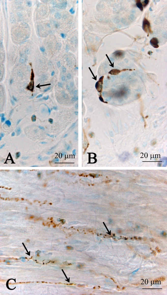
Fig. 1.

CXCL14 immunoreactive endocrine cells in mucosal epithelial cell layer (A, B) and fibers in muscular layer (C) in the columnar epithelial regions of the stomach. Sagittal (A) and transverse (B) sections obtained from the villi. Note that these cells appear to be the closed-type of endocrine cells. Arrows in A and B indicate CXCL14 immunoreactive cells, and arrows in C indicate CXCL14 immunoreactive nerve fibers.
