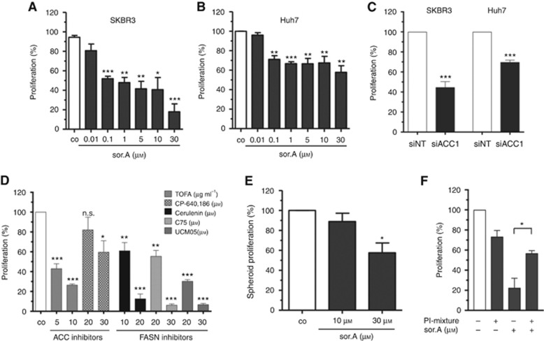
Figure 4.
Abrogated proliferative potential. (A) SKBR3 and (B) Huh7 were treated with increasing concentrations of soraphen A for 96 h and the growth rate was assessed by CellTiter-Blue cell viability assay. (C) Cells were transfected with siACC1 or non-targeting siRNA (siNT), respectively. Proliferation of SKBR3 and HuH7 cells was determined after 96 h. (D) Proliferation of SKBR3 cells treated with different compounds targeting either ACC1 or FASN was determined. (E) Growth of Huh7 spheroids seeded in poly-HEMA plates after stimulation for 96 h. (F) Cells were treated with 10 μM soraphen A for 24 h, then 50 μM of a phosphatidylinositol-mixture was added for further 16 h. Medium was changed and proliferation of the cells was investigated after additional 48 h using CellTiter-Blue reagent. Statistical analysis was performed using Student’s t test: n.s. = non-significant, *P<0.05, **P<0.01, ***P<0.001.

