Figure 1.
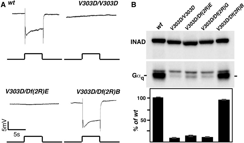
A new Gαq mutant with a flat ERG response. (A) ERG recording in various genetic backgrounds. Flies that are either homozygous for the V303D mutation or trans-heterozygous for V303D and a chromosomal deficiency uncovering the Gαq region “Df(2R)E” (abbreviated for Df(2R)Exel7121) show a nearly complete loss of response to light stimulation. However, flies trans-heterozygous for V303D and a chromosomal deficiency uncovering an adjacent region to Gαq “Df(2R)B” (abbreviated for Df(2R)BSC485) displayed a normal ERG recoding. For all ERG recordings, event markers represent 5-sec orange light pulses, and scale bar for the vertical axis is 5 mV. (B) The level of Gαq protein in various genetic backgrounds. Western blot was used to detect Gαq protein level in whole exact from fly heads with the indicated genotypes. “Df(2R)G” is the abbreviation for Df(2R)Gαq1.3. In each genotype, the Gαq band is marked and the upper band is nonspecific. INAD was used as a loading control. Quantification of the Western blot results is shown below. The complete genotypes are as follows: w1118 (wt); w1118; GαqV303D (V303D); w1118; GαqV303D/Df(2R)Exel7121 (V303D/Df(2R)E); w1118; GαqV303D/Df(2R)Gαq1.3 (V303D/Df(2R)G); w1118; GαqV303D/Df(2R)BSC485 (V303D/Df(2R)B).

