Figure 2.
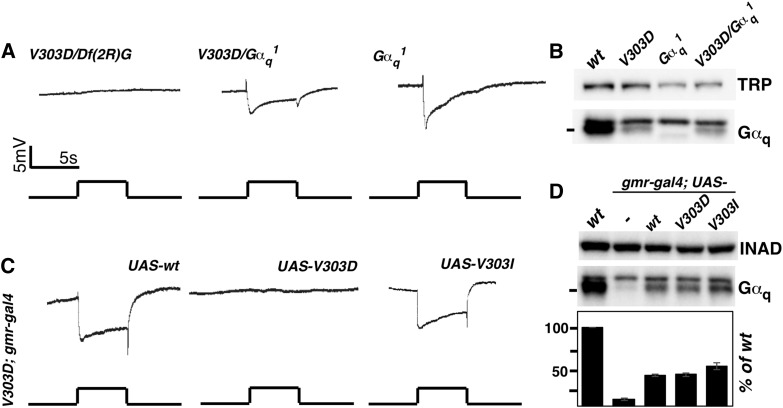
Defective Gαq protein but not the reduction in Gαq level is responsible for the loss of a light response. (A) ERG recordings of Gαq mutants. Flies trans-heterozygous for V303D and the deficiency Df(2R)Gαq1.3 displayed no light response. Mutants either homozygous for the Gαq1 mutation or trans-heterozygous for Gαq1 and V303D displayed a substantial response to light. (B) Western blot analyses of Gαq protein level showed that Gαq level is lower in Gαq1 mutants than in V303D homozygous mutants. TRP serves as a loading control. (C) The ERG recordings of V303D mutants expressing different Gαq variants. Flies carrying homozygous V303D mutation, a GMR-Gal4 transgene, and different UAS-Gαq transgenes were subject to ERG recording. Both the wild-type Gαq and the mammalian mimic V303I transgenes rescued the ERG phenotype. For all ERG traces, event markers represent 5-sec orange light pulses, and scale bars are 5 mV. (D) Western blot measurement of Gαq protein level in rescued lines. Gαq level was restored to 40% of the wild-type level when GMR-Gal4 was used to drive Gαq expression. INAD served as a loading control. Quantification of the Western blot results is given below. The complete genotypes are as follows: w1118 (wt); w1118; GαqV303D (V303D); w1118; GαqV303D/Df(2R)Gαq1.3 (V303D/Df(2R)G); w1118; Gαq1 (Gαq1); w1118; GαqV303D/Gαq1 (V303D/Gαq1); w1118; GαqV303D gmr-Gal4; UAS-Gαq+; w1118; GαqV303D gmr-Gal4; UAS-GαqV303D; w1118; GαqV303D gmr-Gal4; UAS-GαqV303I.

