
Figure 1.
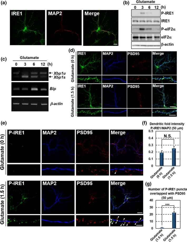
Glutamate stimulation induces local activation of the IRE1‐XBP1 pathway in distal dendrites. Primary cultured mouse hippocampal neurons maintained for 10 days were used for the analyses. (a) Immunofluorescence staining analysis of IRE1 (green) and MAP2 (dendritic marker: red). IRE1 was localized to MAP2‐positive dendrites (Bar: 20 μm). (b) Western blot analysis of phosphorylated IRE1 (P‐IRE1), IRE1, phosphorylated eIF2α (P‐eIF2α) and eIF2α. Cells were treated with 7.5 μM glutamate for the indicated times. P‐IRE1 and P‐eIF2α were transiently up‐regulated following glutamate stimulation. (c) RT‐PCR analysis of Xbp1 and Bip. Spliced Xbp1 (Xbp1s) and Bip were transiently induced by treatment with 7.5 μM glutamate for the indicated times (Xbp1u: unspliced Xbp1). (d) Immunofluorescence staining analysis of IRE1 (green), MAP2 (blue) and PSD95 (postsynaptic marker: red). IRE1 was observed at MAP2‐positive neurites and PSD95‐positive postsynaptic sites of dendrites. The localization and level were not significantly changed by the treatment with 7.5 μM glutamate for 1.5 h. The lower panels show high magnification of the dendrites shown in the upper panels. (Bar in upper panels: 20 μm, in lower panels: 5 μm). (e) Immunofluorescence staining analysis of P‐IRE1 (green), MAP2 (blue) and PSD95 (red). IRE1 was phosphorylated at PSD95‐positive postsynaptic sites of dendrites in response to 7.5 μM glutamate exposure for 1.5 h. The lower panels show high magnification of the dendrites shown in the upper panels. Arrow heads indicate immunoreactivities of P‐IRE1 overlapped with those of PSD95. (Bar in upper panels: 20 μm, in lower panels: 5 μm). (f) P‐IRE1 fluorescence intensities 40–90 μm from the somata of the dendrites in (e). The fluorescence intensities in these dendrites were not increased by glutamate treatment (mean ± SD, N = 10; number of cells prepared from five independent cultures). (g) The number of P‐IRE1‐positive puncta overlapping with PSD95‐positive postsynaptic sites 40–90 μm from the somata in (e). The number of P‐IRE1‐positive postsynaptic sites was increased by glutamate exposure (mean ± SD, N = 10; number of cells prepared from five independent cultures; ***p < 0.001).