
Figure 4.
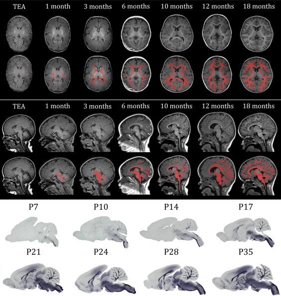
Overview of myelination throughout development from term‐equivalent age (TEA) to 18 months in human infants and from postnatal day (P)7 to P35 in rats. The upper panel shows transverse sections of T1‐weighted MRI scans at different ages, in the lower sections the myelinated white matter is manually colorized (red). The middle panel shows the sagittal sections of T1‐weighted MRI scans at different ages, in the lower sections the myelinated white matter is manually colorized (red). The lower panel shows sagittal sections of rat brains at different ages, stained for myelin basic protein (MBP), a myelin marker. The gross spatio‐temporal pattern of myelination in humans shows high resemblance with that of rodents