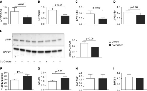
Figure 3.
ASMC phenotype is altered after coculture with epithelial cells. ASMCs were cocultured with BEAS-2B cells for 24 hours. mRNA was extracted to perform RT-qPCR, examining myocardin (MYOCD) (A), α-smooth muscle actin (ACTA2) (B), calponin (CNN-1) (C), and myosin light-chain kinase (MYLK) (D). Open bars, control ASMCs; solid bars, cocultured ASMCs. Protein lysate was separated in a polyacrylamide gel before transfer to PVDF membrane and blotted for α-smooth muscle actin (α-SMA) protein. Control ASMCs: lanes 1, 3, 5, and 7; BEAS-2B cocultured ASMCs: lanes 2, 4, 6, 8 (E). Densitometry of α-SMA blotting normalized to GAPDH is shown. Rate of proliferation was measured by bromo-deoxyuridine (BrdU) incorporation for 18 hours before the end of 24-hour coculture with BEAS-2B cells (F). mRNA for ETS domain-containing protein Elk-1 (Elk1) (G), Kruppel-like factor 4 (KLF4) (H), and serum response factor (SRF) (I) was measured within ASMCs after 24 hours of coculture with BEAS-2B cells. Data are presented as means (+SE). Paired t test was used to compare samples with P values reported above the bars.

