Figure 2.
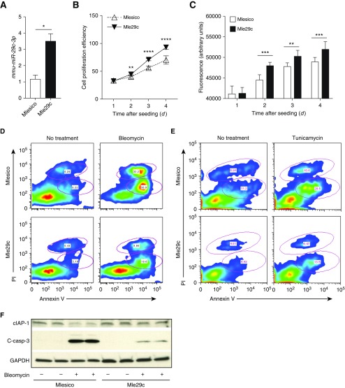
miR-29c overexpression promotes epithelial cell proliferation and inhibits apoptosis. (A) Verification of miR-29c overexpression in stable mouse epithelial cell line tested by quantitative RT (qRT)-PCR. (B) Cell proliferation assay for mouse lung epithelial control cells (Mlesico) and mouse lung epithelial pre-mmu-miR29c over-expressing cells (Mle29c) in medium containing 10% FBS. (C) Viability of Mlesico and Mle29c cells after 4 days of growth in 10% FBS medium. (D) Representative flow cytometry analysis of apoptotic and necrotic cells gated from Mlesico and Mle29c epithelial cells with or without bleomycin treatment (250 ng/ml). (E) Representative flow cytometry analysis of apoptotic and necrotic cells from Mlesico and Mle29c cells with or without tunicamycin (5 ng/ml) treatment. (F) Representative Western blot analysis for cellular inhibitor of apoptosis 1 (cIAP1) and cleaved-caspase (c-casp)-3 in Mlesico and Mle29c cells with or without bleomycin (125 ng/ml) treatment. Representative of three independent experiments (n = 5). *P ≤ 0.05, **P ≤ 0.01, ***P ≤ 0.001, ****P ≤ 0.0001, by Student’s t test (A) or two-way ANOVA (B and C). Data presented are means (±SEM). PI, propidium iodide.

