Figure 1.

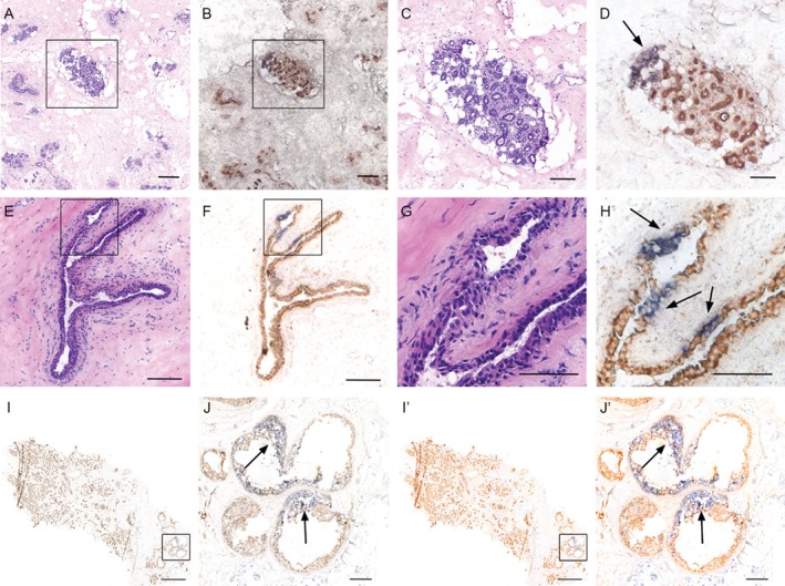
CCO‐deficient patches of cells are found through the normal and premalignant human breast. (A) H&E staining showing a TDLU in the normal adult breast. (B) CCO enzyme histochemistry identifies a subset of cells within the TDLU containing blue, CCO‐deficient cells. High‐power images are shown in C and D, respectively. CCO deficiency is indicated by arrows. CCO‐deficient ducts are also found in the ducts of normal human breast. (E) H&E staining showing a normal duct from adult human breast. (F) CCO enzyme histochemistry identifies three similarly distinct clusters of cells within the normal duct containing blue, CCO‐deficient cells. High‐power images are shown in G and H, respectively. Scale bar = 150 μm; inset scale bar = 75 μm. (I) (and outlined area in J) CCO enzyme histochemistry of a sample of invasive breast cancer with adjacent areas of DCIS identifies a large area of CCO‐deficient blue cells within the premalignant lesion. CCO‐deficient cells are interspersed with wild‐type CCO‐positive brown cells, indicating dynamic mixing of clones in DCIS. Scale bar (I) = 2000 μm; inset scale bar (J) = 250 μm. I' and J' represent globally saturated images (saturation set to 60) to highlight the CCO‐deficient areas in I and J, respectively.
