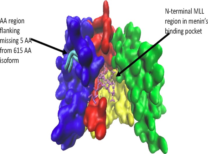
Figure 1.
A 610-amino acid menin isoform (type 2). Note how overall binding groove appears intact and the missing five amino acids are located on the surface away from menin’s binding pocket. This image was rendered from the online RCSB Protein Data Bank code 4GQ6 using VMD 1.9.3 Graphics (University of Illinois at Urbana-Champaign).

